
|
|

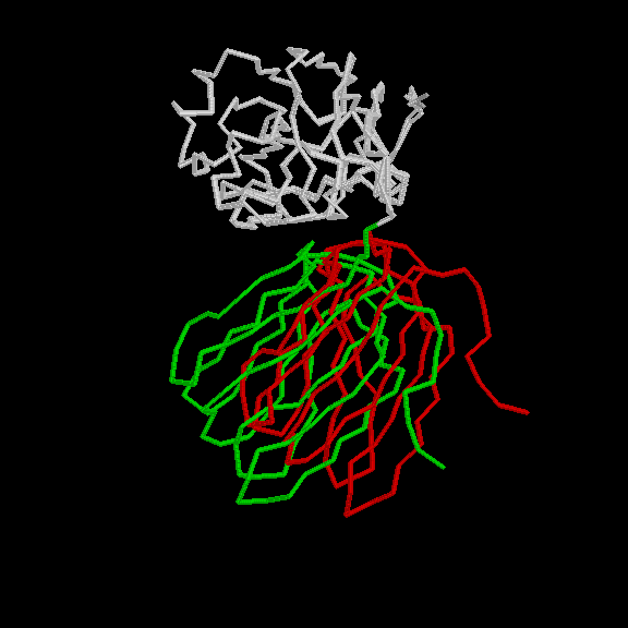
Motion in Tomato Bushy Stunt Virus (TBSV) Coat Protein [tbsv]
[ jump to morphs ]
Classification Known Domain Motion, Hinge Mechanism [D-h-2]
Structures 2TBV Common part starts at residue 101C [ PartsList ] 2TBV Common part starts at residue 101A [ PartsList ]
Description 1 interdomain linkage,1 hinge, ~22 degree rotation. Motion is evident in comparing different subunits in the asymmetric unit. Single data bank identifier applies for both forms. This spherical virus contains 180 subunits arranged with icosahedral symmetry on a T=3 lattice. Each subunit, in turn, contains two major domains, the shell (S) and projection (P) domains that are linked by a peptide in an extended conformation. The symmetry of the virus requires each subunit to fit into one of 3 different packing environments. One of the principal mechanisms for accommodating the different environments is a relative movement of the two domains by ~22 degrees. This movement involves a simple hinge in the peptide connecting the S and P domains.
Particular values describing motion Creation Date = 19970822 Modification Date = 19970822 Annotation Level (1..10) = 7 Location of a Hinge (residue selection) = 266-272 Maximum CA displacement (A) = 13.8 Maximum Rotation (degrees) = 21.6 Experimental Methods = x (Traditional x-ray)
References A J Olson, G Bricogne and S C Harrison (1983). Structure of Tomato Bushy Stunt Virus: The Virus Particle at 2.9 Å Resolution. J. Mol. Biol. 171: 61. [Medline info for 93376766]
Data and Graphics Graphic-1 Graphic courtesy of E S Huang. Torsion Changes + Atom deviations Columns are, respectively: residue, phi-A (-A mean in A subunit), psi-A, sidechain-rotamer-state-A, phi-C, psi-C, rotamer-C, dphi, dpsi, dCA (after doing a fit). Detailed README Describes motion and gives orientation matrices. 4x4 Rotation Matrix The following 4x4 matrix [1 .. 16] orients the opened form so that the axis of rotation is along the z-axis and the origin is at the molecular centroid. MOVIES A page giving pointers to movies of the motion in TBSV.
GO terms associated with structures Molecular function structural molecule activity Cellular component viral capsid
Morphs
[ show all images ]
Best representative Morph Morph name Structure #1 Structure #2 Residues [ ] [ ]
![[help]](http://www0.molmovdb.org/images/help-icon.gif)
![[home]](http://www0.molmovdb.org/images/home-icon.gif)
![[movies]](http://www0.molmovdb.org/images/icon.movie.gif)
Copyright 1995-2005 M. Gerstein, W. Krebs, S. Flores, N. Echols, and others
Email: Mark.Gerstein _at_ yale.edu
/sicon.png)